
Gallery


Zainah Siddiqi Art
"Rampant" is an oil painting focusing on the cell to cell communication and deterioration of the lungs when a person is infected by SARS-CoV-2. The background illustrates how people from all walks of life were impacted by the pandemic.

Varvara Folimonova Art
These four drawings illustrate different types of placentas; epitheliochorial, synepitheliochorial, endotheliochorial, and hemochorial.

Varvara Folimonova Art
These four drawings illustrate different types of placentas; epitheliochorial, synepitheliochorial, endotheliochorial, and hemochorial.

Varvara Folimonova Art
These four drawings illustrate different types of placentas; epitheliochorial, synepitheliochorial, endotheliochorial, and hemochorial.

Varvara Folimonova Art
These four drawings illustrate different types of placentas; epitheliochorial, synepitheliochorial, endotheliochorial, and hemochorial.

Varvara Folimonova Art
These four drawings illustrate different types of placentas; epitheliochorial, synepitheliochorial, endotheliochorial, and hemochorial.

Shanay Desai Art
My summer artwork was focused on creating cover art for a publication/future presentations. Specifically, there is a lack of imagery relating to understanding atherosclerosis and how monocytes induce pro-inflammatory responses. My role was to create a cover art graphic that will represent the inside cross-section of an artery that shows plaque formation and monocyte differentiation between 2 types of monocytes used in lab.

Rina Shou Art
In both abstract art pieces, an amyloid beta plaque touches a microglia cell, which activates a signaling pathway that is involved in the removal of neurotoxic agents. Eventually, the microglia eats the plaque, which limits the progress of Alzheimer's Disease.

Rina Shou Art
In both abstract art pieces, an amyloid beta plaque touches a microglia cell, which activates a signaling pathway that is involved in the removal of neurotoxic agents. Eventually, the microglia eats the plaque, which limits the progress of Alzheimer's Disease.

Rachel Eom Art
An abstract rendering of the intersection between the renal (kidney), cardiovascular (heart), and immune (cytokines shown as white spheres) systems and how changes in the molecular machinery by DNA missense mutation, as well as changes in protein structure greatly influence hypertension conditions.

Pinar Caglayan Art
My artwork consists of modular PNG drawings that aim to explain the MERFISH technology in a simple and clear way. Using these images, I put together a presentation that could be used by Dr. Christina Baer’s lab when introducing this technique to an audience that has no prior exposure to MERFISH.

Nishita Maknojia Art
This image showcases a healthy, normal brain besides an Alzheimer’s brain. The magnifying glass zooms into the diseased brain to highlight how microglia plays a role in AD pathogenesis by releasing inflammatory mediators which can contribute to amyloid beta plaque aggregation.

Nishita Maknojia Art
An abstract image that represents the novel immune intracellular signaling pathway and how activating this immune pathway can function to limit the spread of damaging amyloid beta aggregates in the brain.

Natalie Elliott Art
Unavailable

Nadia Kafil Art
This piece depicts B-cells, antibodies, and strands of free-flowing DNA. These objects are intertwined with one another to represent the connection of these elements in the process of B-cell genome modification and the formation of unique antibodies.

Nadia Kafil Art
This piece depicts a B-cell with its DNA spilling out along with a wave of antibodies flowing through. The piece places a heavy emphasis on the DNA and antibodies of the B-cell which are arranged in a crisscrossing formation to represent the intersection of B-cell genome modification and its role in creating distinct antibodies.

Martina D’Orso Art
The logo I created for Dr. Madhur’s lab is a sleek, contemporary logo that reflects all of their areas of studies combined with the words “Madhur Lab” to easily convey the name and meaning of the lab.

Mariana Smith Art
This dynamic sculpture illustrates the 3D distribution of CcmA proteins on the cell wall surfaces of four H. pylori mutants (with each mutant type shown in a different color under natural light). When black light is introduced, the CcmA protein distribution on each cell becomes visible and begins to glow. Each cell shape is taken from a real cell in the lab, along with its corresponding protein distribution.

Laurence Gao Art
Unavailable

Kritika Bisht Art
This artwork was created to be a logo for the urology-nephrology-hematology teaching grant. It depicts the three branches working together to promote training, mentorship, and collaboration in the form of a happy blood drop. The hematology is represented by the blood drop, nephrology is represented by the lungs, and urology is shown attached to the lungs.

Kim Ly Art
An animation personifying the irony that even though Covid-19 infects the lungs, it is the lungs’ response that triggers severe sickness for the body.

Katelyn Schumacher Art
Logo for the Cassat Lab to represent the laboratory's focus on osteomyelitis research. Depicts principles that are foundational to osteoclast resorption/osteoblast repair processes, while utilizing Vanderbilt Colors.

Katelyn Schumacher Art
Representation of the Cassat Lab's mass spectrometry diagnostic process, as well as the resultant 3D rendering of the infected bone. Serves as a metaphor of how the groundbreaking process is a "light" to the biology community.

Justin Yu Art
Displays the mechanism of the ENaC-Mediated Salt Response, which plays a role in salt-sensitive hypertension.

Justin Yu Art
Displays the overall pathway for salt-sensitive hypertension, showing the organs and tissues involved.

Justin Yu Art
Cover art for a manuscript submitted to the Circulation Research journal, which showcases the article’s focus on salt-sensitive hypertension and the role of inflammasomes. A salt shaker is seen suspended and pouring its contents onto a kidney and blood vessels, representing the tissues that mediate salt-sensitive hypertension. The flames represent the inflammation, while the embers are shaped as inflammasomes and lymphocytes.

John Lee Art
The Lemur Leap frog infected with the chytrid fungus Batrachochytrium dendrobatidis (Bd) has a ready defense in the dermal granular glands. The glands release a steady stream of antimicrobial peptides that can effectively kill the pathogen on the surface of the skin.

Jiawei (Janney) Wang Art
An illustration of the Multiplex Image Labeling With Regional Morphology (MILWRM) pipeline for consensus tissue domain detection across samples in Multiplex Immunofluorescence and Spatial Transcriptomics data.

Grace (River) Terrell Art
This piece shows the distinct effects in PC3 levels based on COVID severity and that infected pregnant women usually had a less severe case of COVID. The final element shows the larger cytokine storm and upregulation of angiogenic factors in non-pregnant individuals.

Gayathree Gopi Art
My first piece depicts the formation of a special type of polymicrobial biofilm called a “mini-biofilm” between C. albicans and the anaerobic bacteria C. perfringens. The second piece illustrates the life cycle and process of mature mini-biofilm formation.

Gayathree Gopi Art
My first piece depicts the formation of a special type of polymicrobial biofilm called a “mini-biofilm” between C. albicans and the anaerobic bacteria C. perfringens. The second piece illustrates the life cycle and process of mature mini-biofilm formation.

Erin Lee Art
The task was to create a urology-nephrology-hematology teaching grant logo. It was requested that the interaction between the three branches were highlighted, along with an emphasis on collaboration, mentorship, training, and diversity.
The goal was achieved by showing the different branches on separate slides and having different hands come together to create the final illustration.

Erika Noda Art
Unavailable

Ereny Morcos Art
This summer, I worked in Dr. Nardhy Gomez-Lopez's lab to help generate a representation of the maternal-fetal crosstalk in humans and mice using single-cell technologies. My digital artwork showed the results received from a study the lab conducted where single-cell RNA-sequencing revealed unique cellular interactions in preterm labor that was driven by intra-amniotic infection.

Emily Krueger Art
My piece is an animation on the progress of a Group B Strep infection through the fetal membrane in pregnancy. This infection and resulting inflammation of the fetal membrane can have dangerous consequences during pregnancy, including premature labor and possible infection of the fetus.

Elaina Lewis Art
This analogy represents contact networks as bubbles that leak viral particles. It describes how contact networks are not as small as they are perceived, and viruses can be spread unknowingly outside of the bubble.

Elaina Lewis Art
This represents the spread of viral particles in a population through contact networks and cryptic transmissions. As pathogens with different epidemiological parameters move through the population, an increase in contacts results in more infection and spread.

Elaina Lewis Art
This visualizes a concept for a global city with famous landmarks from around the world. It is a comment on how globalization results in an increase in the spread of viruses throughout the population.

Diana Espinoza Art
This poster is meant to highlight the necessity of having compassion during stressful times such as a major health crisis. Part of what I believe contributes to having a better understanding of how a pandemic affects our peers and society is through obtaining efficient data visualization and modeling which the Colubri lab has been dedicated in making more accessible through multidisciplinary means.

Diana Espinoza Art
This abstract work is meant to depict the rapidly evolving nature of mRNA viruses. The work contains motifs related to the replication of viruses in the body which are then used to compose a scene that mimics that of a city in chaos.

Ardria McDonald Art
It represents how B cells develop, divide, and branch out to other organs from bone marrow, in which it all resembles a growing “tree”.

Ardria McDonald Art
It depicts DNA transcription, as well as antigens to show that they are very closely related in the process of transcription and translation. Transcription of Antibody coding genes drives somatic hypermutation and antibody diversity

Anna Mehlhorn Art
In order to help audience members conceptualize sexual biofilms as they relate to conventional biofilms, I created a figure displaying both in the fungal species, Candida albicans. I intentionally used a soft color palette and cartoon-like illustrations to make an intimidating concept more approachable to the viewer.

Alyssa Glauser Art
A diagram identifying the layers of the fetal membrane and how they come into contact with the maternal blood and decidua.

Aimee Li Art
My contributions involved a poster and two handouts. The microscope poster was designed for middle school students especially affected by COVID-19. Both the handouts are related to a brain activity for the same middle schoolers.

Aimee Li Art
My contributions involved a poster and two handouts. The microscope poster was designed for middle school students especially affected by COVID-19. Both the handouts are related to a brain activity for the same middle schoolers.

Aimee Li Art
My contributions involved a poster and two handouts. The microscope poster was designed for middle school students especially affected by COVID-19. Both the handouts are related to a brain activity for the same middle schoolers.

Zhizhu Zhang Art
This cartoon depicts a spaceship delivering the LTB4 lipids, using the actual microscopic image taken by the Serezani lab as background. The inspiration of spaceship was taken from an interesting mistake of the computer vision technology. When Dr. Serezani sent over some microscopic pictures for reference, Gmail described the pictures as "[A picture containing star, outdoor object, night sky, light Description automatically generated with low confidence]". Produced by macrophages and neutrophils, LTB4 is an important signaling mediator to enhance antimicrobial effector functions. However, excessive LTB4 can lead to aberrant inflammation that is damaging to host defense. Thus, some LTB4 molecules are portrayed to be devil-like.

Zhizhu Zhang Art
This cartoon depicts the immune cells and bacteria during abscess formation. Some neutrophils grab the magenta-colored S. aureus as a representation of phagocytosis. Others shoot arrows at the bacteria, which shows the process of exocytosis/release of ROS and proteases. Some dead neutrophils are also contained within the abscess, undergoing NETosis to trap the bacteria as well. The abscess is then encapsulated with fibrous and macrophages, which is represented by macrophages holding the barricade tapes at the perimeter. The cartoon uses the actual microscopic of the abscess formation process in the background. Zhizhu hopes to capture the dynamic interaction between different host immune components and invading microbes while making science fun and accessible.

Varvara Folimonova Art
Activation-induced cytidine deaminase (AID) is an essential element within B cells for generating antibodies, which help to defend the human body against infections.

Varvara Folimonova Art
A topologically associating domain (TAD) is a region within the genome where genes are more likely to interact with one another. Dr. Basu's lab studies TADs along with other genetic elements within B cells to better understand their mechanism of generating antibodies.

Varvara Folimonova Art
A topologically associating domain (TAD) is a region within the genome where genes are more likely to interact with one another. Dr. Basu's lab studies TADs along with other genetic elements within B cells to better understand their mechanism of generating antibodies.

Shubhanjali Minhas Art
This cover art was created for a CRISPR/Cas9 system that the Nobile lab was working on. The piece likens a block tower to the genome of Candida auris, a fungus highly resistant to antifungal treatment. A Cas9 molecule is represented in the piece along with guide RNA. When this Cas9 molecule is used to take out certain genes, represented by individual blocks, in the genome, the block tower eventually falls, representing the heightened susceptibility of Candida auris to antifungal treatment. The light blue glow in the background represents the nucleus of Candida auris cells, while the orange globular shapes in the back represent the Candida auris cells.

Shubhanjali Minhas Art
This is another version of the CRISPR/Cas9 cover art. Here, the block tower is represented in a "zoomed in" environment, where a monochromatic blue color scheme is used to represent this event occurring in the nucleus. The background contains many strands of dna, further illustrating that this is occurring in the nucleus of a Candida auris cell.

Rebecca Dubin Art
This picture shows how the COVID-19 molecule is engulfed by a cell. The ones and zeros represent the computational aspect of the lab which is important to interpret and organize results and to uncover new discoveries.

Rebecca Dubin Art
This image depicts a concept that the Denison lab has been working on. One aspect that makes COVID-19 difficult to stop is that it has a proofreading mechanism protecting the replication of its RNA known as ExoN. It has been discovered that although 5FU, a common drug used to cause mutations, is blocked by ExoN, a drug known as Remdesivir is able to sneak around the ExoN bouncer.

Rebecca Dubin Art
This piece shows how science meets the real world effects of COVID-19. The lungs represent the experience of many people who become ill with the virus. Inside the lungs there is a COVID-19 molecule with a microscope on the left and the replication complex of Corona-virus RNA on the right.

Rebecca Dubin Art
This work shows the concept of coronavirus spillover. It is a pinball machine showing several different species that have been shown to carry coronaviruses. The coronavirus ball travels around hitting the different species and earning points for the virus.

Qi (Kathy) Liu Art
Using her "luminescent" art style, Kathy depicts three possible approaches to HIV-1 vaccine development. Six types of trimers from six strands of HIV-1 are indicated using different colors. The three approaches from top to bottom are the mixture of trimers, DNA nanoparticles with six different trimers attached, and groups of three 2-component insect ferritin with two trimers attached to each. She also created artworks for the three approaches respectively in a similar art style, making them a set of artworks for vaccine development.

Michelle Kwon Art
Acinetobacter baumannii is a unique bacteria that tends to be widespread in hospital ICUs and surface tops. Rather than becoming weaker without the presence of water, it actually expresses the protein DtpA which allows the bacteria to tolerate environmental conditions without water. This piece depicts the bacteria in the center, glowing, as it expresses its unique protein while being surrounded by a body of water. The water that wraps around the bacteria is distant from the body representing the strength of the bacteria even without the direct presence of water. Furthermore, the blue water-like structure plays a dual role as it also represents the folded DtpA protein structure. Surrounding this entire figure are other bacterias that would usually need water and the presence of moisture in order to survive.

Melina Woods Art
Unavailable

Melina Woods Art
This tree of life shows the relationship between different fungal species that form chlamydospores.

Lynette Butron Art
This image replaces human lungs with Cryptococcus spores.

Lynette Butron Art
This is a rendering of an MRI scan that replaces the hemispheres of the brain with Cryptococcus spores. This relates to Cryptococcus' connection with meningitis.

Lynette Butron Art
This image depicts a macrophage engulfing Cryptococcus spores.

Lucy Britto Art
Comparison of the normal function of iron handling receptor CD136 and how metabolic dysfunction may impact CD163 shedding mechanisms such as enzymatic cleavage or extracellular vesicles.

Lucy Britto Art
Comparison between the normal function of iron handling receptor Transferrin (TfR1) and the iron overload condition that has been implicated in decreased soluble TfR1, increased TfR1 saturation, and transcriptional dysfunction.

Lucy Britto Art
Describes the normal function and mechanism of ferroportin, a multi-cellular iron exporter, compared to the influence of metabolic disease that may impact iron recycling via increased transporter degradation or mutations.

Lucy Britto Art
Overview of the relationship between iron homeostasis and the iron responsive elements/iron regulatory protein (IRE/IRP) system that coordinates the uptake, export, and storage of iron through post-transcriptional control mechanisms.

Lucy Britto Art
Depiction of the normal role of the heme oxygenase-1 protein in the heme handling pathway and how its function or ablation plays multiple regulatory roles in glucose and lipid homeostasis in a variety of cells and tissues.

Lucy Britto Art
Visual schematic of a 17-day experimental protocol performed by a postdoc in the Hasty Lab involving primary cell isolation to study to the transfer of iron between differentiated adipocytes and polarized macrophages.

Kadeer Wellington Art
This piece captures the stepwise assembly of a herpesvirus virion from the perspective of an assembly manual. The viral capsid is assembled in the nucleus of infected cells and is composed of distinct parts. This component will be combined with the viral genome. The second complex is prepared within the cytoplasm consisting of a lipid envelope containing viral proteins. The final steps involve combining the assembled capsid with the assembled viral lipid envelope to produce an infectious particle. Each part is digitally drawn and edited using MediBang Paint Pro.

Justin Yu Art
This is a logo created for the Karijolich Lab. It includes a dark bar running through its center that is supposed to represent the black resin tops of lab benches. As seen in the logo, lab equipment commonly used in biomedical laboratories sits on top of this black bar. In the lower half, the "o" in Karijolich has been replaced with an icosahedron containing dsDNA, a representation of the Kaposi's Sarcoma-associated Virus, an important focus for the Karijolich Lab. Arrayed around the letters are a number of viral RNA structures that the lab studies as well.

Justin Yu Art
This piece of cover art depicts an anthropomorphized virion wearing a suit with a head shaped like an icosahedron, representing the Kaposi's Sarcoma-associated Virus (KSHV), a major focus of the Karijolich Lab. Jutting out from the back of the virion are a number of different mechanical elements, which are used to signify the synthetic nature of viruses, as they rely on a host's cellular machinery in order to reproduce. One gear is being removed by a disembodied hand. This represents the Karijolich Lab's research into the many mechanisms of KSHV infection, including the relationship between ORF36 and ISGylation.

Justin Edaugal Art
This digital piece illustrates the David Lab's research on Graft-versus-Host Disease (GVHD). A probiotic was given to a mouse model to study its affects on microbe behavior in the gut. This drawing is inspired by a cross-sectional, microscopic image of a normal colon on the left, and an image of an inflamed colon on the right as well as the atomic structures involved in the research.

Justin Edaugal Art
A mixed media piece representing concepts of nutrition and bacteria. Portrait of a Filipino farmer holding a basket of fruit, representing nutrition, made using graphite pencil on paper. The portrait was then cut and layered with colored paper beneath to represent microbial dynamics. The Filipino farmer portrait is a nod to Filipino heritage of the David Lab Principal Investigator and the artist, myself. Graphite pencil, colored pencil on cut and layered, paper.

Justin Edaugal Art
This digital piece illustrates a graphic comic-style, representation of the David Lab's research. What we eat can affect the concentration and type of microbes in our gut over time. Digital media.

Justin Edaugal Art
Inspired by the shapes and movement of microbes and tissue cells of the human gut, this digital piece relates the dynamics of microorganisms with the dynamic movement of water, each being dependent on their environments. Digital media.

Justin Edaugal Art
This piece is an abstract interpretation of the David Lab's research. The David Lab is interested in researching how nutrition affects microbial dynamics in the human gut over time. This piece combines colors and shapes from a microbial stream plot with a cross-sectional, microscopic image of an intestine. Colored pencil on black paper.

Helen Qian Art
A depiction of drug molecules binding onto the CFTR protein to remedy loss of function mutations in cystic fibrosis.

Helen Qian Art
A depiction of organelles in action on the folding and trafficking pathway of the cystic fibrosis transmembrane conductance regulator (CFTR) protein.

Gina Yu Art
Working with SARS-Cov-2 is much like taking apart a complex puzzle and piecing it back together. Unable to work with the virus directly, techniques such as cloning the RNA into a yeast cell to form a Yeast Artificial Chromosome (YAC) have been used to then selectively target for mutations with eukaryotic Multiplex Automated Genome Editing, or eMAGE. The ability to manipulate this positive-strand virus in such a manner is what allows us to piece together how this virus behaves.

Ereny Morcos Art
Unavailable

Emma Thomas Art
This is a graphical abstract that shows an uninfected neural organoid with a rosette on the left, and a HCMV infected neural organoid with a disrupted rosette on the right. The information in the middle conveys what processes and cells are either down regulated, absent, or unaffected with the HCMV infection.
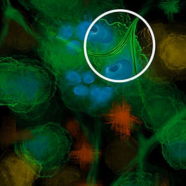
Emma Thomas Art
This is an abstract piece conveying the idea of syncytia. The green represents cells and the blue is representing nuclei. The idea of syncytia is that many cells fuse together into one large multinucleotic cell. The white circle offers a point of focus and distinction where the haziness of the abstract comes into more focus and detail.

Elsa Runquist Art
This is a diagram describing the different techniques used by the Lopez Lab when studying copy-back defective viral genomes (cbDVGs). These techniques include VODKA, RNA FISH, and PCR. All three techniques are mapped out on a single page, allowing for greater efficiency when delivering this information to other students and researchers. This illustration was initially made in BioRender, and then it was replicated using Photoshop drawing tools. (This is the Photoshop version).

Elsa Runquist Art
This is a schema describing the process for how copy-back defective viral genomes (cbDVGs) are formed and their impact on the physiology of the cell, along with the relationship to respiratory syncytial virus (RSV) disease severity in infants. This illustration was initially made in BioRender, and then it was replicated using Photoshop drawing tools. (This is the Photoshop version).

Ellen Yu Art
Three conditions are commonly associated with diabetic retinopathy: high blood pressure, high fat, and high glucose. These conditions are represented by the chemical structures of angiotensin, palmitic acid, and glucose respectively. These factors impacted gene expression, so I inserted them inside loops of DNA, similar to the function of histones. This led to phenotypic changes in mueller glia, astrocytes, and microglia resulting in an overall phenotype that closely resembles diabetic retinopathy.

Elaine Lewis Art
This is an acrylic painting that shows a complementary color scheme. It represents one of the Lopez Lab's main area of study: respiratory viruses.

Elaine Lewis Art
This is a digital representation of the heterogeneity of amounts of defective viral genomes (DVGs) and full viral genomes in different cells during respiratory viral infection. The cells have different functions if there are a large number of DVGs in a cell compared to full viral genomes.

Elaine Lewis Art
This is a digital schema of the different pathways defective viral genomes (DVGs) can undergo as they are integrated into a cell. DVGs can cause viral replication interference with the full viral genome. They could also interact with PKR and cause stress granules and antiviral immunity. Finally, they could undergo the RLR/MAVS signaling pathway and cause cell survival/persistence, antiviral immunity, or inflammation.